
Training courses
The University of Manchester at Harwell (UoMaH) is bringing the imaging community together by delivering courses covering a broad range of topics to help support researchers using Harwell’s national facilities.
Recognising the need for advanced training skills in the Harwell Campus, a network of academic and industrial partners from UoMaH, 3Dmagination, Diamond, ISIS, and Finden have jointly developed training courses at different levels to meet this need with an emphasis on data analysis.
The current course topics cover:
- fundamentals of X-ray tomography data visualisation, AI-assisted analysis and quantification
- advanced scripting for batch processing of tomographic data
- 4D tomography with digital volume correlation (DVC)
- correlative imaging
If there is a course you would like to see us offer, please get in touch, and we will see what we can do!
Current courses available
A 2 day training course covering theory and computer-based practical works using the Thermo Scientific™ Amira-Avizo Software.
Lab/synchrotron X-ray tomography has emerged as one of the most important techniques for research in a wide range of applications including healthcare, energy, food and geology. Advances in the quality of X-ray beams, optics, high speed data acquisition and in-situ environments have made significant improvements to non-destructive imaging of a specimen structure in 3D and over a wide range of length scales (micron- and nanoscales). Although the direct benefit of X-ray tomography is the 3D visualisation of the internal structure of specimens, which is very valuable to understand the structure/function relationships, the technique has however a lot more to offer. The information is hidden in the data, and robust methods and workflows — increasingly powered by AI/deep learning — are needed to extract it in a relevant and accurate way, and make it readily available for decision making.
Organisation
The training is organised in three parts: theory of image processing and deep learning (0.5 day), and computer-based practical work using the Thermo Scientific™ Amira-Avizo Software 3D (1.5 day), including its AI-based tools for denoising and segmentation. At the end of the second day, the users are invited to practise on their own dataset with the help of the trainers. The attendees are invited to bring their own laptop (an Avizo license will be provided for the training).
Who should attend?
This course is aimed at both beginners with no prior experience in 3D imaging and image processing, and intermediate-level researchers who want to know more about it and explore different ways of analysing 3D datasets, including the latest AI-assisted workflows.
Learning outcomes
- Basics of image processing and mathematical morphology
- Introduction to the theory of Deep Learning: what neural networks are and how they learn, convolutional neural networks (CNNs) and why they suit image data, how a network is trained (data, labels, loss, epochs), and the practical implications for tomographic data (e.g. data/label quality, generalisation, and when AI methods are a better fit than classical ones)
- Import/manipulate tomographic dataset
- Surface/volume renderings
- Filtering
- AI-based denoising of noisy/low-dose tomographic data
- Basic/advanced segmentation
- AI-based segmentation, including 2D and 2.5D deep learning approaches
- Quantification
- Animation/movies
How to register
Please contact Sarah Batts if you wish to sign up for this course: sarah.batts@manchester.ac.uk. Places are limited to 10 attendees per course.
Price
Academics, students, postdocs, government: £400.00 + VAT. Industry: £800.00 + VAT.
Although X-ray tomography is an attractive characterisation technique in materials science, it generates a huge amount of data at a fast rate and it can be extremely time consuming to process the data manually. A great benefit to creating scripts and plugins in Avizo is the ability to reuse a workflow on more than one image or to develop a bespoke workflow for more complex materials.
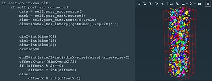
The training is organised in two parts: Python scripting in Avizo (1 day) and development of Avizo Python module (1 day). At the end of the second day, the users are invited to practise on their own dataset with the help of the trainers. The attendees are invited to bring their own laptop (an Avizo license will be provided for the training).
Who should attend?
This course is aimed at advanced users with prior expertise in 3D imaging and image processing of 3D datasets. Prior experience with Avizo and/or Python is recommended.
How to register
Please contact Sarah Batts if you wish to sign up for this course: sarah.batts@manchester.ac.uk. Places are limited to 10 attendees per course.
Price
Academics, students, postdocs, government: £400.00 + VAT. Industry: £800.00 + VAT
Digital Volume Correlation (DVC) is a powerful experimental technique that computes 3D full-field displacement and strain maps from volumes images acquired during a deformation process of a material. DVC is the 3D extension of Digital Image Correlation (DIC) which was first described four decades ago. The emergence of DVC started early 2000s with the use of X-ray CT combined with in situ rigs to capture 3D morphological changes with time.
Although the DVC algorithms are in spirit similar to DIC algorithms, DVC requires special attention and expertise from multiple fields (3D imaging, in situ testing, CT reconstruction, mechanics of materials, etc.) as the texture is not controlled (natural contrast brought by imaging) and the noise/CT artefacts are often dominating the accuracy/precision of the measurements.
Adding the 4th dimension (3D + time) also means that data processing and visualization are key to extract the mechanical information hidden inside the 4D datasets.
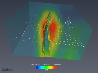
The training is organised in two parts: theory (0.5 day) and computer-based practical works (1.5 day) using the XDigitalVolumeCorrelation extension in the Thermo Scientific™ Amira-Avizo Software 3D where two solutions are implemented (a classic subset-based approach and a more robust global approach). At the end of the second day, the users are invited to practise on their own dataset with the help of the trainers.
Who should attend?
Researchers working in the field of mechanics, physics, and materials science using 3D imaging techniques such as X-ray computed tomography who are interested to (i) compute 3D full-field displacements and strains for linking the microstructure to the mechanical/physical behaviour, (ii) use the DVC measured data to feed/validate finite element models. The attendees are requested to bring their own laptop to practise on the XDigitalVolumeCorrelation extension (free trial licence will be provided).
How to register
Please contact Sarah Batts if you wish to sign up for this course: sarah.batts@manchester.ac.uk. Places are limited to 10 attendees per course.
Price
Academics, students, postdocs, government: £400.00 + VAT. Industry: £800.00 + VAT.
General course information
Correlative imaging combines data from multiple acquisition techniques and length scales to build a more complete picture of a specimen than any single modality can provide on its own. This is particularly powerful when combining 3D X-ray or neutron tomography with high-resolution 2D or 3D techniques such as FIB-SEM, allowing researchers to link bulk-scale structural context with fine, local microstructural detail. However, correlating datasets acquired at different resolutions, on different instruments, and sometimes with different contrast mechanisms, introduces a range of practical challenges: alignment, registration, normalisation, and interpretation. This course covers the acquisition principles and the practical Avizo-based workflows needed to bring these datasets together reliably.
Organisation
The training is organised in two parts: theory (0.5 day), covering an introduction to acquisition techniques and the principles of correlative workflows, and computer-based practical work using the Thermo Scientific™ Amira-Avizo Software 3D (1.5 day). At the end of the course, users are invited to practise on their own dataset with the help of the trainers. The attendees are invited to bring their own laptop (an Avizo license will be provided for the training).
Prerequisites
Prior attendance of a foundational tomography/image processing course (e.g. “Visualisation, AI-Assisted Analysis and Quantification of Tomographic Datasets”) is recommended, or equivalent working experience with 3D image processing.
Who should attend?
This course is aimed at researchers who already have some familiarity with 3D image processing and who want to combine multiple imaging modalities and/or resolutions in their analysis workflow. It is particularly relevant to those working with FIB-SEM, multi-scale tomography, neutron/X-ray correlative studies, or any workflow requiring registration of datasets from different instruments.
Learning outcomes
Introduction to acquisition techniques:
- Overview of X-ray tomography (lab and synchrotron) and its resolution/field-of-view trade-offs.
- Overview of neutron tomography: complementary contrast mechanisms to X-ray (e.g. sensitivity to light elements/hydrogenous materials), typical facilities, and where it adds value in a correlative study.
- Overview of FIB-SEM acquisition: serial sectioning principles, imaging modes, and typical artefacts.
- Other correlative modalities (e.g. SEM/EDS, confocal, other 2D imaging) and where they fit into a correlative workflow.
- Understanding contrast mechanisms across modalities and their implications for registration and interpretation.
FIB-SEM data processing:
- Import and inspection of FIB-SEM image stacks.
- Alignment correction of serial sections (drift, curtaining, and misalignment between slices)
- Tilt correction.
- Grayscale normalisation across slices and datasets.
Working with multi-resolution and multi-modality data:
- Handling datasets of different resolutions within the same project.
- Resampling strategies and their impact on data fidelity.
- Registration of datasets from different modalities (rigid and non-rigid approaches), including X-ray/neutron tomography pairs and tomography/FIB-SEM pairs.
- Defining and extracting regions of interest for correlative analysis (e.g. locating a FIB-SEM ROI within a lower-resolution tomography volume).
- Quality-checking and visualising correlative results.
Practical workflows:
- End-to-end example: from lower-resolution tomography to FIB-SEM ROI, aligned and registered.
- Illustration of quantification from multiple modalities.
- Working on your own dataset with trainer support.